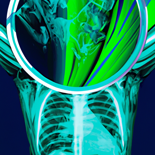
The Discovery of X-Rays: Revolutionizing Medical Imaging

The Discovery of X-Rays: Revolutionizing Medical Imaging
The discovery of X-rays in 1895 by German physicist Wilhelm Conrad Roentgen has had a profound impact on the field of medicine. Not only did it revolutionize medical imaging, but it also opened doors to new avenues of exploration in various scientific disciplines.
Roentgen's accidental discovery occurred while he was experimenting with cathode rays. He noticed that a fluorescent screen placed nearby emitted a bright glow even when covered with a black paper. Roentgen deduced that there must be a new kind of ray that was causing this fluorescence, and he named it X-ray, as "X" represents an unknown variable.

Once Roentgen discovered X-rays, he began experimenting and collecting evidence on their properties. He found that X-rays can penetrate through materials that are opaque to visible light, such as human tissue, clothing, and even metal. This unique property led to the realization that X-rays could be utilized for medical imaging.
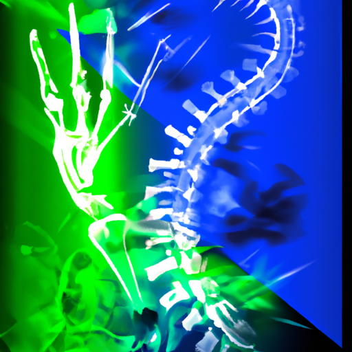
Roentgen's initial experiments involved taking X-ray images of objects, including his wife's hand. He was able to produce an image that showed the skeletal structure of her hand, highlighting bones and other dense structures. This breakthrough led to the realization that X-rays had the potential to revolutionize medical diagnoses by allowing doctors to visualize the internal structures of the human body without invasive procedures.
One of the earliest medical applications of X-rays was in dentistry. Dentists could now examine teeth and the surrounding bone structure to identify issues such as cavities, fractures, and impacted teeth. This new imaging technique played a crucial role in improving dental care and diagnosis.
The use of X-rays expanded rapidly in medicine, and it became especially valuable in orthopedics. X-rays could now help doctors assess fractured bones, identify dislocations, and determine the severity of injuries. This allowed for more accurate diagnoses, thereby improving treatment outcomes for patients.

Another significant contribution of X-rays to medical imaging was the identification of tumors and cancerous growths. X-rays could provide insights into the location, size, and characteristics of tumors, aiding physicians in determining the appropriate course of action. X-ray mammography, for instance, has become an indispensable tool in detecting breast cancer at an early stage.
X-rays also played a vital role in surgical procedures. Utilizing X-ray imaging during surgery allowed surgeons to navigate intricate anatomical structures, such as blood vessels and nerves, with greater precision. This led to improved surgical outcomes and minimized complications.
Over time, advancements in technology have further enhanced the effectiveness and safety of X-ray imaging. Digital X-ray machines have replaced traditional film-based methods, allowing for quicker image acquisition and manipulation while reducing radiation exposure to patients. Additionally, computed tomography (CT), a technique that employs X-rays in multiple angles to obtain highly detailed cross-sectional images, has revolutionized medical imaging even further.
The discovery of X-rays by Wilhelm Conrad Roentgen has undoubtedly revolutionized medical imaging and transformed the field of medicine. Through X-ray technology, doctors gained a non-invasive and safe method to see through the human body, leading to earlier and more accurate diagnoses. As technology continues to advance, it is likely that X-rays will continue to evolve, improving patient care and expanding the boundaries of medical knowledge.


